Cryostats are among the most important pieces of equipment in dermatology and histology labs. If you’ve ever wondered how dermatologists can diagnose skin cancer the same day they remove tissue, or how Mohs surgery achieves such high cure rates, the answer is often the cryostat.
This guide is designed for beginners — whether you’re a medical assistant, lab technician, or simply curious about how these machines work. By the end, you’ll understand what a cryostat is, how it operates, and why it’s so vital in dermatology.
What Is a Cryostat?
A cryostat is a machine that freezes and slices thin sections of biological tissue for examination under a microscope. Think of it as a highly specialized, refrigerated cutting tool. Unlike traditional methods that use paraffin embedding (a process that takes hours or even days), a cryostat makes it possible to prepare slides within minutes.
In dermatology, cryostats are most closely tied to Mohs micrographic surgery, the gold-standard treatment for certain types of skin cancer. During Mohs, tissue is removed, frozen, cut into thin slices, stained, and examined under a microscope — all while the patient waits. The surgeon repeats the process until no cancer cells remain, ensuring maximum precision with minimal healthy tissue loss.
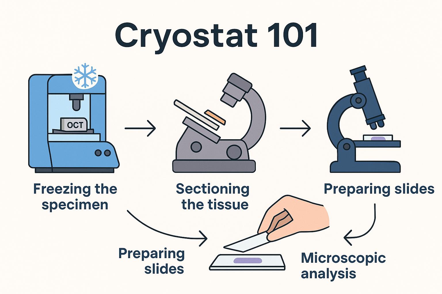
How a Cryostat Works
At its core, a cryostat is both a freezer and a precision slicer. Here’s what happens inside:
- Freezing the Specimen
- The tissue is placed in a medium called OCT (Optimal Cutting Temperature compound) and mounted on a small metal disc.
- This disc attaches to the cryostat’s object head, which is cooled to subzero temperatures (usually between -20°C and -30°C).
- Sectioning the Tissue
- Once frozen solid, the object head moves the tissue block toward a razor-sharp blade.
- The built-in microtome slices the block into sections as thin as 5–10 microns (a fraction of the width of a human hair).
- Preparing Slides
- Each tissue slice is carefully placed on a glass slide.
- Slides are then stained (often with hematoxylin and eosin, or H&E) so cells are visible under a microscope.
- Microscopic Analysis
- The surgeon or pathologist examines the slides immediately.
- If cancer cells are still visible at the margin, another thin layer of tissue is removed from the patient, and the cycle repeats.
The Role of Cryostats in Dermatology
Cryostats have applications across medical fields, but in dermatology they are most essential for:
- Mohs Surgery – Enables same-day margin control for skin cancer.
- Histology Labs – Used to prepare frozen sections for diagnosing skin diseases and conditions beyond cancer.
- Research – Dermatology researchers use cryostats to study tissue architecture and cellular structures.
The advantage of cryostats in dermatology is speed and precision. By bypassing slower paraffin embedding techniques, doctors can make real-time treatment decisions without sending samples to an outside lab.
What Using a Cryostat Looks Like
For beginners, it helps to picture the workflow step by step. Imagine you’re assisting in a Mohs procedure:
- The surgeon removes a small layer of tissue.
- You bring the specimen to the cryostat and embed it in OCT.
- Within minutes, the machine freezes the block.
- Using the handwheel, you advance the tissue block and cut ultra-thin slices with the microtome.
- The slices are transferred to glass slides, stained, and handed back to the surgeon for immediate review.
This process repeats until the surgeon confirms all cancerous cells have been removed.
Key Features of a Cryostat
While different brands and models vary, most cryostats share these essential features:
- Freezing chamber – Keeps tissue at subzero temperatures.
- Object head – Holds and positions the specimen block.
- Microtome blade – Slices frozen tissue with high precision.
- Anti-roll plate – Prevents tissue sections from curling as they’re cut.
- Controls and display – Allow users to set temperature, thickness, and other cutting parameters.
Together, these features make cryostats both powerful and user-friendly once you’re trained.
Why Beginners Should Learn Cryostat Basics
Understanding cryostats is important even if you’re not the one operating them daily. For dermatology staff, knowing the basics helps you:
- Appreciate why Mohs surgery can be done in one visit.
- Communicate more clearly with patients about the process.
- Support surgeons and lab technicians during high-volume procedures.
- Recognize the value of routine training and proper workflow.
Cryostats are not just lab equipment — they’re a critical part of how modern dermatology delivers faster, more precise care.
What You Need to Know Before Operating a Cryostat
Operating a cryostat takes training and care. Before stepping up to one, beginners should keep these key points in mind:
- Temperature Control – Cryostats operate between -20°C and -30°C. Always confirm the chamber has reached the correct temperature before sectioning.
- Specimen Embedding – Tissue must be embedded in OCT compound and frozen solid before cutting. Poor embedding leads to uneven or unusable sections.
- Blade Safety – The microtome blade is extremely sharp. Use caution when loading, adjusting, or cleaning around it. Always engage safety guards when not cutting.
- Anti-Roll Plate Use – To keep tissue slices flat, make sure the anti-roll plate is positioned correctly above the blade.
- Section Thickness – Most dermatology sections are cut at 5–10 microns. Always set thickness according to the procedure (e.g., Mohs vs. routine histology).
- Slide Handling – Frozen tissue is delicate. Use clean, dry slides and handle them gently to avoid damaging sections.
- Staining Workflow – Know the basic staining process (commonly H&E) so slides can be prepared promptly for the surgeon or pathologist.
- Defrosting – Cryostats build up frost over time. Learn when and how to perform defrost cycles to keep the chamber working efficiently.
- Cleaning – Daily cleaning of the chamber and blade holder prevents contamination and keeps sections clear.
- PPE & Hygiene – Always wear gloves, lab coats, and eye protection. Tissue samples are biohazardous, and the cryostat chamber is a controlled environment.
These fundamentals aren’t troubleshooting steps — they’re the baseline knowledge every beginner should have before operating a cryostat in a dermatology setting.
Final Thoughts
Cryostats may look intimidating at first glance, but at their core they are simply machines designed to do one thing very well: freeze and cut tissue into thin slices for immediate analysis.
For dermatology practices, they are indispensable. By enabling real-time tissue examination during Mohs surgery, cryostats give doctors the ability to remove cancer with pinpoint accuracy while preserving healthy skin.
For beginners, the takeaway is this: a cryostat is the bridge between surgery and diagnosis, and mastering its role is key to understanding modern dermatology workflows.